Description:
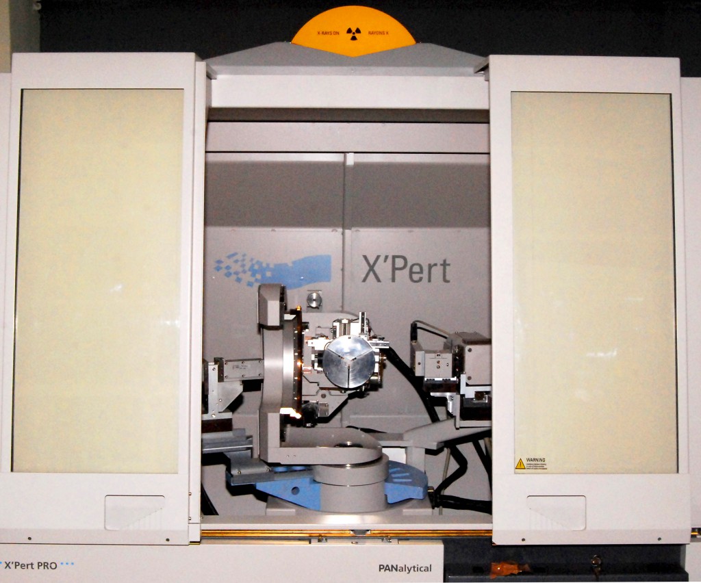
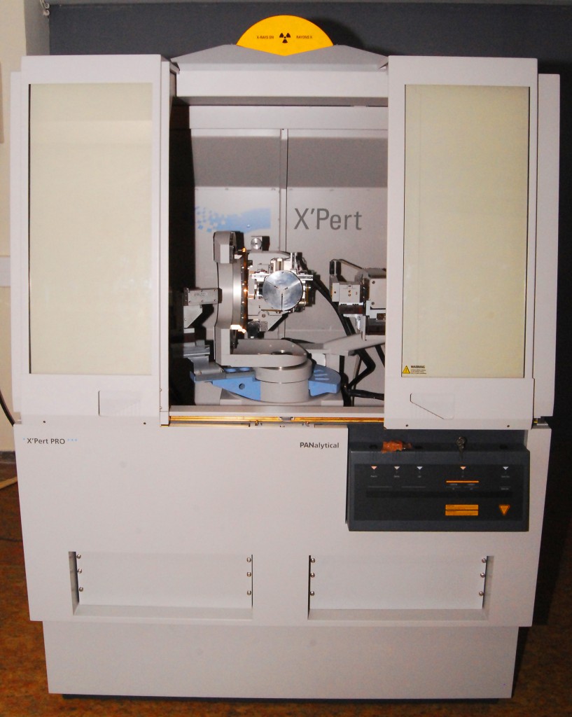
The Panalytical X’Pert Diffractometer is a highly advanced, versatile materials characterization system. Interchangeable PreFIX incident and diffracted beam optics can be configured for optimal measurement of high resolution scans, reflectivity experiments, or for in-plane diffraction.
High Resolution X-ray Diffraction: High resolution x-ray diffraction is generally applied to highly ordered crystals. It is useful to examine nearly lattice matched materials or the structural perfection of materials. The system can be used to accurately measure the width of Bragg diffraction peaks from nearly perfect single crystals over the range of a few seconds of arc. In order to obtain the high resolution, the angular divergence of the incident x-ray beam must be very small with little or no peak broadening due to spectral dispersion and the goniometer must be capable of very accurate stepping. Special incident and diffracted beam conditioning makes this possible. This system can also map regions in reciprocal space around the Bragg reflections which can be useful to characterization relaxation of strained epitaxial films.
Reflectometry: Reflectometry is an analytical technique for investigating thin layers. In reflectivity experiments, the X-ray reflection of a sample is measured around the critical angle. Below the critical angle of total external reflection, X-rays penetrate only a few nanometers into the sample. Above this angle the penetration depth increases rapidly. At every interface where the electron density changes, a part of the X-ray beam is reflected. The interference of these partially reflected X-ray beams creates the oscillation pattern observed in reflectivity experiments. From these reflectivity curves, layer parameters such as thickness and density, interface and surface roughness can be determined through modeling.
In-plane diffraction: In-plane diffraction is a diffraction technique in which both the incident and diffracted beams are nearly parallel to the sample surface. Because the beam is incident at a grazing angle, the penetration depth of the beam is limited to within about 100 nm of the surface. The in-plane diffraction technique measures diffracted beams nearly parallel to the sample surface and hence measures lattice planes that are (nearly) perpendicular to the sample surface. These planes are inaccessible by other techniques.
Equipment:
- Selectable line or point focus 1.8kW sealed ceramic copper x-ray tube source.
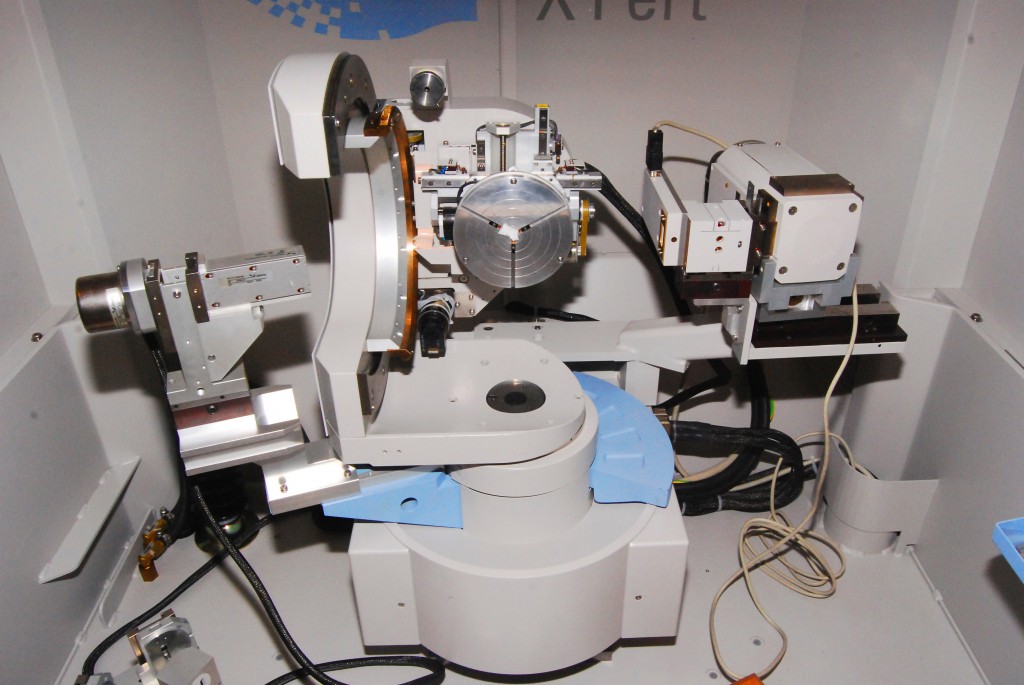
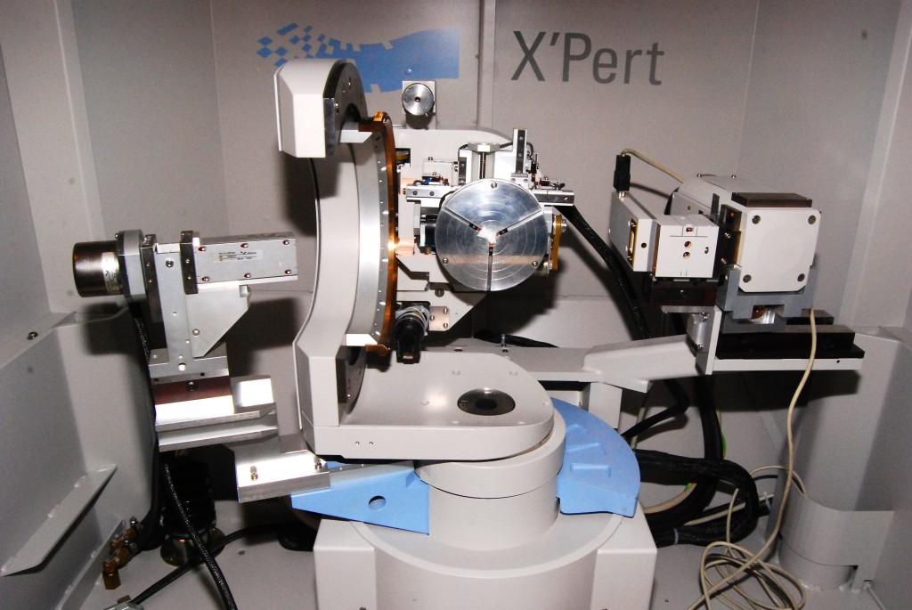
- Choice of PreFIX incident beam optics include a graded parabolic x-ray mirror with automatic attenuator, four-bounce Ge(220) monochromator, hybrid four-bounce monochromator with mirror and automatic attenuator, fixed divergence slits, or x-ray lens.
- High resolution goniometer with optically encoded sample positioning enables a minimum step size of 0.0001°.
- 1/2 circle Eulerian cradle with motorized sample stage enables sample tilts of +/- 90°, in-plane rotation of 360°, in-plane X and Y translations of 100 mm, and vertical Z displacement of 11 mm.
- Sample holder accommodates samples up to 4 inches in diameter.
- Choice of PreFIX diffracted beam optics include triple axis/rocking curve optics or parallel plate collimator.
- Triple axis setup utilizes a three bounce (022) channel cut Ge crystal to provide an acceptance angle of 12 arc seconds.
- The rocking curve optics utilize interchangeable slits to control the background and detector resolution.
- The parallel plate collimator has a 0.27° acceptance angle with an optional 0.1 mm collimator slit to improve the resolution at 2Θ angles less than 4°.
- 2 sealed proportional detectors with a large dynamic range.
 |
 |
 |
 |
Accessories:
- Thin-Film Optics
Applications:
- High Resolution
- Rocking curves.
- Superlattice scans.
- Reciprocal space maps.
- Substrate offcut.
- Epitaxial layer tilt.
- Layer relaxation.
- Epitaxial layer mismatch.
- Reflectometry
- Composition analysis structure of grain boundaries in ceramics.
- Magnetic films on chromium.
- Identification of precipitates in materials.
- Analysis of nanoparticle sizes.
