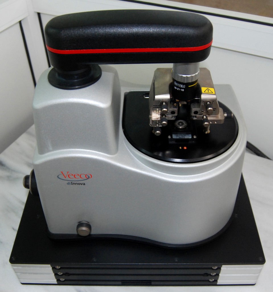
Description:
The Veeco diInnova AFM is capable of measuring surface topography.- Large area scans (XY = 90um) where Z = 7.5um.
- Small area scans (XY = 5um) where Z = 1.5um.
Imaging Modes:
- Contact Mode: This is the most basic and straightforward AFM mode. In this case, the AFM probe tip is in constant physical contact with the sample surface.
- Tapping Mode: The most commonly used AFM mode for imaging soft or fragile samples. This technique maps topography by lightly tapping the sample surface with an oscillating probe tip.
- Phase imaging: This is a secondary imaging mode derived from tapping mode that detects variations in composition, adhesion, friction, viscoelasticity, electric and magnetic properties. Phase imaging maps the phase lag between the periodic AFM cantilever oscillations.
Specialized Operations:
- Lateral Force Microscopy: This is derived from contact mode by measuring the lateral bending of the cantilever. Information regarding the friction characteristics of a sample can be determined.
- AFM Force Spectroscopy: Provides force-distance curve measurements by record the amount of force felt by the cantilever as the probe tip is brought close to and even indented into a sample surface and then pulled away. This technique can elucidate local chemical and mechanical properties like adhesion and elasticity e.t.c.
- Electrostatic Force Microscopy (EFM): Measures electrical field gradient distribution above the sample surface. EFM relies on LiftMode, a two-pass technique. During the "first pass" topography of the image is recorded in tapping mode and surface plane parameters are obtain. During the "second pass" a voltage is applied to AFM tip which scans the surface, with the same scanning parameters, at a designated height recording long-range electrostatic interactions. EFM is used for electrical failure analysis, detecting trapped charges, mapping electric polarization, and performing electrical read/write, among other applications.

